2016年7月28日 星期四

Am I a bioinformatician ?

當我再次回到這個 blog 時,有點感到遺憾。

本來有點想要維持每週一篇,看來還是很難的。

我想,這裡可以分享一些經驗談,不是每個人都要走類似的過程,不過,或許會有人受惠於這些經驗的。

前一陣子,或是每隔一陣子,就會有人好奇的問我,怎麼從生物學訓練的背景轉變做生物資訊研究?我應該說的是,我一直沒有很認真的走上生物資訊研究,只是潮流和趨勢,現在已經回頭不易了。

跨領域嗎?沒人認真的這樣看待,就這樣,自認作了一些覺得有意義的研究,是好事吧。

第一步?我會這樣說,人生的契機。我在很早的時候,在沒有 xml 的定義之前,就「會」html。怎麼學會?自學。自學,所以在世界上越來越多資訊背景的人開始加入制訂各種定義與標準之後,我就不像以前那麼「會」,因為我完全沒有科班背景。不過我很容易懂別人的想法,所以在入門時,會比純實驗室背景的人快。

每當身旁從事生物學研究的朋友問起時,我總有點心虛,因為我不是寫 code 的人,不過我可以看得懂怎麼用,專心投入多一點的時候,有時還可以看得懂人家怎麼寫,就改寫成自己需要的。在有 google 大神的神助下,我的世界也就這樣地運行。但是:

​我只記得我需要知道的部分。

這樣講或許不負責,或是不知恥,但是這個世界有那麼多需要知道的事情,google 一天收集的資料量有多少?這些怎麼能夠全部裝在我的小小不能加 硬碟/CPU/RAM 的那個腦裡面?腦科學家說,腦裡面的 synaps 連結方式的複雜如同宇宙一般,所以我們可以做那麼多的事情。只可惜我以前自豪的強大記憶力已經漸漸衰落,上週才跑過的程式,我現在就已經忘光了!那些參數怎麼設定,沒有筆記或是 log (翻譯:留存的記錄檔),我完全不能重複這些程序。

簡單來說,現在我的腦海裡還是留存著很多在分子生物實驗室長期訓練中所學會的實驗程序,然而我也一樣,即使在當下,在我腦力還蠻驚人的時候,我仍然不會倚賴我所記得的細節。哪一個 reagent 要加多少 ul ? 這種細節不應當憑靠著記憶,而是參照 protocol 來完成。用同樣的原理來說,執行某種分析所使用的程式可能有幾種,每種的參數給法都不大一樣,所以,我只記得我需要完成的程序,但細節就要看 menu。

你同意嗎?這是經驗法則戰勝教室的證據嗎?或者是考選與訓練被放大過多?


嗯 ~~~~~  不需要考據的作文寫起來真快。

2016年4月11日 星期一

Ho Ho Ho.....Hox.


Hox Genes in Development: The Hox Code

By: PZ Myers, Ph.D. (University of Minnesota, Morris) © 2008 Nature Education
http://scienceblogs.com/pharyngula/2007/09/the_hox_code.php


What is Hox genes?

  • Hox genes, a family of transcription factors, are major regulators of animal development. Unlike most genes, however, the order of Hox genes in the genome actually holds meaning.  These are genes that specify segment identity; the expression pattern is translated into the many different forms that animals (metazoans) exhibit.
  • Discovery of Hox: Drosophila study
    果蠅有8個 hox genes (the top array of genes in figure 1) / 1 個 hox cluster。實驗發現這些基因異常時會造成體節構造異常:例如,Antp ==> Antennapedia。

Genomic organization of the Hox gene cluster (Figure 1 on the source article).  ** 這個圖的方向要把左邊看成3',基因的排列方向(sense strand/ translation)等於要從右向左。zen, bcd, ftz  是 非 hox 的 homeobox containing genes. 
  • 比較果蠅和小鼠的hox 構造,有幾個明顯的不同:
    • number of hox casettes: 4 vs 1
    • 5' 與腹部發育有關的基因,小鼠多好幾個 (hox9-13 vs Abd-B)
  • 這個基因與cluster 數量的差距,顯然可以讓我們聯想到[產生比較多樣的組合來控制基因]===> 構造多樣化,"hox code" ?
    • 果蠅:某個 segment 表現某一種 hox (不會有兩個 hox 都表現
    • 小鼠:某個 segment 表現一個以上 (同位的)hox ( hox4 可達四個)==> 魚可以有八個?
  • multiple copies of hox 或許也是有功能上的 rescue 好處,例如 HoxA3 (表現在頸椎與頭相接的部位)ko 並沒有明顯表型/功能缺失,推測為 HoxD3 的補償作用。只做 D3 mutant 時,第一節頸椎出現嚴重的構造缺失,但還是有頸椎。double mutant A3, D3 則變成這節與頭部癒合==> 顯示 HoxA3 果然還是決定第一節頸椎構造的關鍵;構成這節骨頭的中胚層起始組織可受到相當程度的重新限定分化,而癒合到顱骨的構造去。( 原文:HoxA3 is important after all; without it, the first neck vertebra doesn't form. In fact, in this instance, it is thought that the initial mesodermal tissue for the bone is so thoroughly respecified that it fuses completely with the skull instead, becoming part of the base of the skull). 
  • 要產生正確脊椎構造,需要適當的hox 基因表現組合,在小鼠的基因剔除分析的實驗結果分析變得相當複雜,即使是很重要的基因,只剔除一個基因可能顯現不出什麼表型缺陷,而必須把整群 "paralogous" 基因都剔除。這也是為什麼脊椎動物的 homeotic mutation 不像果蠅那麼容易被觀察到。
  • In flies, one gene can be mutated, resulting in an altere (平衡桿,退化的第二對翅膀)being transformed into a wing, or an antenna turning into a leg; in the mouse, two to four genes must be simultaneously removed to get a similar complete transformation.)

Paralogous Knockouts in Mice

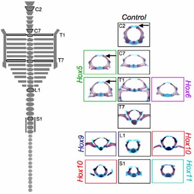
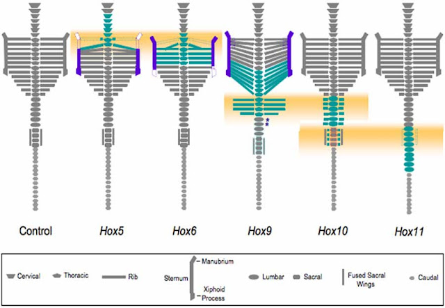
技術上的演進-小鼠發育生物學家發表了一些把整個 paralogous hox 拿掉的實驗結果: (figure 2, dorsal view and concludings in figure 3)
  • delete all Hox5 genes: make C7 and T1 stay like C2 (cervical vertebrae with no dorsal neural arch) and T1 with an incomplete develop ribs.
  • delete all Hox6: no ribs at T1; (almost) a complete homeotic transformation of T1 to C7.
  • delete Hox9: extra ribs at L1: anterior shift in the midtrunk (extend the segment or shorter tail?)
  • delete Hox10: extra ribs at L1~S1
  • delete Hox11: no fused sacral wings at S1    


Changes in specific vertebral elements for the Hox5, Hox6, Hox9, Hox10, and Hox11 paralogous mutants  (figure 2 in the source).


Schematic representation of regions of reported phenotypes in Hox paralogous mutants. (figure 3 in the source).


Schematic of overlaps in and differences between the somite-derived primaxial phenotypes and the lateral plate-derived, abaxial(離軸) phenotypes of Hox paralogous mutants.

(remarks: from p261, ch16, "Bones and Cartilage: Developmental and Evolutionary Skeletal Biology" by Brian K. Hall. DOI: http://dx.doi.org/10.1016/B978-0-12-416678-3.00016-1)

  • primaxial : structures that differentiate entirely within the somitic environment- the vertebral column, vertebral ribs, periaxial and intercostal uscles and associated connective tissue - and that are generated entirely from somitic cells
  • abaxial: all structures that differentiate within the lateral plate environment, including the migrating somite-derived myoblasts that migrate into and differentiate within lateral-plate mesoderm, namely limb and abdominal muscles, sternal muscle and sternum.)

每一個 Hox paralog 控制某特定型態,不僅是由 paralog 內的組合,也包含paralog 間的組合。比如說,在薦椎段(sacral segment)有 Hox10 與 Hox11表現。其他的實驗顯示這裡的分化基礎型態為"胸椎",推測 hox10 hox11 的作用為抑制肋骨形成。

所以,脊椎動物的中軸骨骼形成上,看起來彷彿有 ”數位化” 混合 Hox 控制的機制。

=====================================================================
Another material for easy-understanding Hox:

http://learn.genetics.utah.edu/content/variation/hoxgenes/

1. Scientists discovered homeotic genes by studying strange transformations in fruit flies. Scientists called these modifications "homeotic transformations," because one body part seemed to have been replaced by another. 


Top: (Left) Normal fruitfly; (Right) Fruitfly with mutation in antennapedia gene Bottom: (Left) Normal fruitfly; (Right) Fruitfly with a homeotic mutation that gives it two thoraxes. Bottom images courtesy of the Archives, California Institute of Technology and the whole sources are from learn.genetics http://learn.genetics.utah.edu/content/variation/hoxgenes/

It is awsome to view the body domain swayed by hox genes in arthopods:


2016年3月8日 星期二

DNA methylation

In eukaryote and prokaryote world, as a genetic material, DNA molecules stably transferred from generation to generation (a cell to its daughter cells, the parents to their offspring) in terms of the sequence context........ in most of the time.

Genome changes. A special type of modification in the base is "methylation".  DNA methylation draws scientists' eyeballs because it has been proved to play roles in GENE EXPRESSION.  The genome can be changed the activity without change the context.  And some of these marks changed in parents can be transmitted to offsprings (epigenetic effect) 

More than 20 types of epigenetic and DNA-damage modifications have been identified, most scientists have only been able to study one type, cytosine methylation.

===============================================================

Review

Nucleic Acid Modifications in Regulation of Gene Expression

by Kai Chen, Boxuan Simen Zhao, and Chuan He

Cell Chemical Biology, Volume 23, Issue 1, 21 January 2016, Pages 74–85
doi:10.1016/j.chembiol.2015.11.007

========************************************=========

Abstract

Nucleic acids carry a wide range of different chemical modifications. In contrast to previous views that these modifications are static and only play fine-tuning functions, recent research advances paint a much more dynamic picture. Nucleic acids carry diverse modifications and employ these chemical marks to exert essential or critical influences in a variety of cellular processes in eukaryotic organisms. This review covers several nucleic acid modifications that play important regulatory roles in biological systems, especially in regulation of gene expression: 5-methylcytosine (5mC) and its oxidative derivatives, and N6-methyladenine (6mA) in DNA; N6-methyladenosine (m6A), pseudouridine (Ψ), and 5-methylcytidine (m5C) in mRNA and long non-coding RNA. Modifications in other non-coding RNAs, such as tRNA, miRNA, and snRNA, are also briefly summarized. We provide brief historical perspective of the field, and highlight recent progress in identifying diverse nucleic acid modifications and exploring their functions in different organisms. Overall, we believe that work in this field will yield additional layers of both chemical and biological complexity as we continue to uncover functional consequences of known nucleic acid modifications and discover new ones.

========************************************=========

5-Methylcytosine Methylation in Higher Eukaryotes

The existence of cytosine methylation (5mC) in genomic DNA was first reported by Wyatt in 1951 (Wyatt, 1951).


5mC writer:
  • establish 5mC (de novo): Dmnt3a, Dmnt3b, Dmnt3L,
  • maintain C methylation from hemimethylated DNA at CpG site: Dmnt1
5mC effector/ reader, proteins that recognize 5mC and carry out subsequent actions:
  • bind to methyl-CpG: MeCP1,MeCP2, MBD1, MBD2, MBD4.


Scheme of the Reversible Cytosine Methylation in DNA and Binding Proteins that Are Known To or Proposed To Bind Modified Cytosine Derivatives, Derived from Liyanage et al., 2014  (Figure 1. from the reference)


5mC eraser: not a simple action by one or few enzyme:
  • TET proteins are methylcytosine dioxygenase that utilize dioxygen to oxidize 5mC==> 5hmC==>5fC==>5caC
  • Both 5fC and 5caC can be recognized and excised by human thymine DNA glycosylase (TDG), followed by base excision repair (BER) to replace the modified cytosine with a normal cytosine.
  • or, diluted through cell division (5hmC, 5fC, and 5caC ==> to the unmethylated stage) 

DNA methylation modulates the chromatin structure and affects cognate gene expression by maintaining various expression patterns across cell types (Cheng and Blumenthal, 2010 and De Carvalho et al., 2010).

in promoter   ===> suppression;
in gene body ===> possitive correlation with gene expression 


Two models for "Methylation induced gene repression (promoter):
  1. "indirect” model: DNA methylation may recruit its reader proteins that act as transcription repressors, preventing transcriptional factors from accessing the promoter region.
  2. "direct” model: DNA methylation may be a disruptor to interfere with the binding of certain transcription factors and thus prevent the activation of corresponding genes
 ## of course the world is not so simple........... the transcription regulation roles of DNA methylation typically synergize with various histone marks as the methyltransferases, demethylases, and readers of DNA methylation interact with various histone marks or histone modification enzymes.


DNA 5mC methylation was considered to be dynamic and reversible.


N6-methyladenine (6mA) found in Genome


In bacteria, 6mA serves as an important marker participating in DNA repair, replication, and cell defense (restriction–modification (R-M) systems,in which 6mA, 5mC and 4mC can be recognized by corresponding restriction endonucleases as a label to prevent the host genome from restriction digestion and further enable the degradation of unmethylated foreign DNA (Murray, 2002)) .

** 4mC modifications can be differentiated at base resolution using a revised TAB-seq protocol (4mC-Tet-assisted-bisulfite- sequencing, Yu et al., 2015, Nucleic Acids Res., 43, e148).

In addition to prokaryotes, several eukaryotes have relatively abundant 6mA in genomic DNA. In 2015, three groups reported the presence of 6mA in three different eukaryotes (algae chlamydomonas, C elegans, Drosophila ) independently.


C.elegans genome was thought lacking DNA methylation. The discovery of 6mA, its demethylase NMAD-1, and potential methyltransferase DAMT-1 in C elegans changes this view ===> will 6mA serves as DNA methylation mark (instead of 5mC)?


Figure 2.

N6-Methylation on Adenine in Genomic DNA

(A) A brief overview of biological function of methyl groups in bacterial genomic DNA.

(B) High-throughput mapping of N6-methyladenine (6mA) in Chlamydomonas reinhardtii revealed a unique distribution pattern in the genome with complete depletion at transcription start sites (TSS) and high enrichment at the linker region between nucleosomes.

(C) In Caenorhabditis elegans, 6mA is installed by DAMT-1 and reversibly removed by NMAD-1. The “crosstalk” between 6mA and histone modification, particularly the histone H3 methylation, indicates critical roles that 6mA may play in gene expression regulation.

(D) 6mA in Drosophila melanogaster could be converted back to A by Tet homolog DMAD. Intriguingly, the 6mA level is correlated with the expression level of transposon, supporting the regulatory significance of 6mA in eukaryotes.


Evidence of 6mA function in other situation:
  • In zebrafish, the knockdown of METTL3 (mRNA m6A writer) leads to smaller heads, eyes, and brain ventricles, and curved notochords (Ping et al., 2014)
  • m6A is the most prevalent internal modification in mRNAs and long non-coding RNAs (lncRNAs) in higher eukaryotes (Wei et al., 1975).
  • in the mammalian transcriptome, approximately three m6A marks exist per mRNA molecule and occur within a consensus motif of G(m6A)C (70%) or A(m6A)C (30%), but the methylation percentage at each site varies substantially (all from studies before 1990)
for mRNA,
  • m6A “writer:
    a complex of methyltransferase-like 3 (METTL3), methyltransferase-like 14 (METTL14), and Wilms' tumor 1-associating protein (WTAP)
  • m6A “eraser: (AlkB family proteins) the fat mass and obesity-associated protein (FTO) and ALKBH5
  • m6A reader: YTHDF1 and YTHDF2


N6-Methyladenosine (m6A) in mRNA and Its Biological SignificanceThe reversible methylation and demethylation process occurs in the nucleus, catalyzed by methyltransferase complex and demethylases, respectively. The 6mA modification has profound effects on mRNA fate: it switches mRNA to active translation mode, and also accelerates its decay rate. Figure 3 from the reference

pseudouridine (Ψ) =====> epitranscriptomics ?

https://en.wikipedia.org/wiki/Epitranscriptomics

((here is a plenty info about discoverings and function of modified bases in RNA (mRNA, tRNA, rRNA, miRNA........))) ---beyond my interestings now.


Outlooks

  • We now appreciate that DNA methylation, as a bona fide epigenetic marker, is not only inheritable and dynamic, but also involved in diverse regulatory processes.
  • The recent discoveries of 6mA as a functional DNA mark in eukaryotic genomic DNA raise the possibility that 6mA plays regulatory roles complementary to 5mC.
  • With a better understanding of 6mA methyltransferase and 6mA demethylase and discovery of potential reader proteins, DNA methylation looks to be a ubiquitous epigenetic marker in almost all kingdoms of life.
  • internal m6A methylation in mRNA was shown to be reversible


====================================================================

DNA N6-methyladenine: a new epigenetic mark in eukaryotes?

Luo, GZ et al., 2015. Nature Reviews Molecular Cell Biology
16, 705–710  doi:10.1038/nrm4076


  • Most eukaryotic 6mA research has focused on unicellular protists; 6mA accounts for ~0.4–0.8% of the total adenines in these genomes
    • Recently, 6mA was detected by multiple approaches in the genomic DNA of two metazoans, C. elegans (0.01–0.4%) and D. melanogaster (0.001–0.07%)
    • In prokaryotes, most of the 6mA sites are located within palindromic sequences
  • 6mA is the most abundant internal modification in mRNAs and is widely conserved in eukaryotes. 


Figure 1 | Methods to detect N6 -methyladenine (6mA) in genomic DNA.
Left: an antibody against 6mA can sensitively recognize 6mA and enrich 6mA-containing DNA for subsequent next-generation sequencing (NGS); 6mA-sensitive restriction enzymes can specifically recognize either methylated or unmethylated adenines in their recognition-sequence motifs, and this can be captured by sequencing to determine the exact locations of 6mA.
Right: liquid chromatography coupled with tandem mass spectrometry (LC–MS/MS) can differentiate methylated adenine from unmodified adenine and quantify the 6mA/A ratio by normalization to a standard curve; single-molecule real-time (SMRT) sequencing can detect modified nucleotides by measuring the rate of DNA base incorporation (dashed arrow) during sequencing.


2016年2月18日 星期四

Hemoglobins and Hb related genes

雖然是很早就知道,也很早就有人研究,不過,還是重新瞭解一下吧。

Evolution of hemoglobin and its genes

by Hardison RC


Abstract
======================================================================
Insights into the evolution of hemoglobins and their genes are an abundant source of ideas regarding hemoglobin function and regulation of globin gene expression. This article presents the multiple genes and gene families encoding human globins, summarizes major events in the evolution of the hemoglobin gene clusters, and discusses how these studies provide insights into regulation of globin genes. Although the genes in and around the α-like globin gene complex are relatively stable, the β-like globin gene clusters are more dynamic, showing evidence of transposition to a new locus and frequent lineage-specific expansions and deletions. The cis-regulatory modules controlling levels and timing of gene expression are a mix of conserved and lineage-specific DNA, perhaps reflecting evolutionary constraint on core regulatory functions shared broadly in mammals and adaptive fine-tuning in different orders of mammals.
======================================================================
深入探究血紅蛋白(hemoglobins)與其基因的演化,提供我們對血紅蛋白的功能與基因表現調節的豐富題材。本論文主要針對幾種人類 globin 基因與基因家族,歸納血紅蛋白基因簇演化過程中的主要歷程,並討論這些研究如何深入闡明血紅蛋白的調節。座落於 alpha-like globin 基因複合集(α-like globin gene complex)內或周圍的基因相對地比較穩定,而beta-like globin 則有較大的變動,這可能是基因移動到一個新位置的證據,以及常見的族系專有(lineage-specific)的增多或刪減。控制基因表現量與表現時機的順位調節模組(cis-regulatory module)是保守性的與族系特有的DNA 綜合效果,這或許能反映出哺乳類廣泛共有的核心調控功能的演化限制,以及哺乳綱不同目別物種的適應性微調。

What is Hemoglobin ?

  • transport/bind oxygen
  • it is an abundant proteins in red blood cells of mammals and other jawed vertebrates (gnathostomes). 
  • In human, Hb is known as a heterotetramer : two α-globin and two β-globin, each with an associated heme group.


The model of evolution of vertebrate globin genes. Figure 1 of this reference.
  • Myoglobin:  monomeric heme-bound globin protein found predominantly in skeletal and heart muscle.  It has long been described as an oxygen storage protein.
  • Cytoglobin: found in many tissues.
  • neuroglobin: Its mRNA is abundant in brain tissue but also is present in many other tissues. It is related to invertebrate nerve globins.
  • Physiological function of the heme-bearing proteins are "oxygen binding".  But, CYGB showing nitric oxide dioxygenase activity (converting NO to NO3-) and NGB showing nitrite reductase activity to form nitric oxide  (converting NO3- to NO. This catalytic capability also found in MB and deoxy-Hb). Thus, the nitrite reductase activity could provide a means to produce nitric oxide under hypoxic conditions, signaling from which could regulate mitochondrial respiration and protect tissues (nerves by NGB, heart muscle by MB) from damage under ischemic conditions (Dietz 2011). 
  • MB, CYGB, and NGB are present as single-copy genes, whereas HBB and HBAs are in clusters with multiple related genes.
  • cyclostomes (represented by hagfish and lampreys), also use a heme-containing globin for oxygen transport, but surprisingly, it is more closely related to CYGB than to the gnathostome hemoglobins. This suggests that the oxygen transport function of heme-containing globins arose by independent, convergent evolution in the two major branches of vertebrates. (((Is there sure, solid evidences for these conclusions, or just speculation by the authors?)))
  • Human: alpha-, beta-, delta-, tau-, epsilon- globin.  expressed in different locations / different dev stage. The hemoglobins produced at distinct developmental stages have different affinities for oxygen and are subject to complex regulation by cofactors, favoring an overall movement of oxygen from the maternal bloodstream to that of the fetus or embryo.
  • In human, the separation of α-like and β-like globin gene clusters in amniotes requires coordination of expression between different chromosomes. 人類 (有羊膜類)的α-like and β-like globin gene clusters 分別在不同的染色體,需要協同控制表現。
  • Fish species show an interesting contrast, in that the gene cluster orthologous (homologous genes generated by a speciation event) to that of the mammalian α-globin gene cluster contains both α-like and β-like globin genes ((((((the expression level is not like to be in this guess))))))))) 魚類的 α 與 β globins 則在同一個 cluster 中 (MN type) 
    • MN: found in all gnathostomes being examined. flanked on one side by the genes MPGNPRL3
    • LA (or LCA):  The globin genes in this locus are flanked by the genes LCMT1 and AQP8, and the locus can be called “LA.” The gene ARHGAP17 is also part of this locus in many species. These three nonglobin genes are in the same arrangement and order in the tetrapods (human, platypus, chicken, and frog), but the globin genes is devoid
    • DS: DCHS1 on one side and STIM1 on the other; the RRM1 gene is adjacent to STIM1 in many species. In placental mammals, hundreds of OR genes are in this locus, with additional multigene families such as TRIM genes. (DCHS1 and STIM1 are spanned by many many genes==>several megabases away). Found in "amniotes" (water-tighted environment for embryo. reptiles, birds, mammals) but absent in fish and amphibians.
      b-like globin only.  


Models for evolution of hemoglobin gene complexes in jawed vertebrates. Figure 2 from the reference. The gene maps are not complete, nor are they to scale. (This diagram is adapted fromHardison 2008.)


Maps of orthologous α-like globin genes and expression timing in amniotes. Figure 2 from the reference. 

The history of the gene clusters encoding hemoglobins is dynamic and complex.  (((inferred to be a more adaptive to the environment).

The ortholog of the μ-globin gene is expressed in adult erythroid cells in birds, producing αD-globin

Hemoglobin gene expression is restricted to erythroid cells.

===================... contents in the further section discuss the gene regulation, gene origins, ...... are majorly in "mammal", not my interesting subject now.  so I skipped   


2016年2月16日 星期二

choanoglafellate

======================================================================
另一個題外話: choanoglafellate  (wiki : https://en.wikipedia.org/wiki/Choanoflagellate),  a group of free-living unicellular and colonial flagellate eukaryotes considered to be the closest living relatives of the animals.

The cell body of many choanoflagellates is surrounded by a distinguishing extracellular matrix or periplast.

A free living SPERM ? with a Coat/ a reinforced, moving home?


The choanoflagellates feed on bacteria. Movement of the flagellum draws water through the collar, and bacteria and detritus are captured by the microvilli and ingested.Water currents generated by the flagellum also push free-swimming cells along, as in animal sperm.


A model of S. rosetta life history. Fig 2 from "Cell differentiation and morphogenesis in the colony-forming choanoflagellate Salpingoeca rosetta" by Dayel et al., 2011. (doi:10.1016/j.ydbio.2011.06.003


Analysis on molecular phylogenetic analysis on SSU, LSU, a-tubulin and hsp90 found that choanoflagellates are strongly supported as monophyletic and confirm their position as the closest known unicellular living relative of animals.

Two choanoflagellate species have had their genomes fully sequenced, with another two species having had transcriptome data published.


Previously, Choanoflagellida was divided into these three families based on the composition and structure of their periplast; now with classification based on molecular phylogenetics.  The choanocytes (also known as "collared cells") of sponges (considered among the most basal metazoa) have the same basic structure as choanoflagellates. Collared cells are found in other animal groups, such as ribbon worms,[17] suggesting this was the morphology of their last common ancestor.


The SIT gene family shows little or no homology to any other genes, even to genes in non-siliceous choanoflagellates or stramenopiles. This suggests that the SIT gene family evolved via a lateral gene transfer event between Acanthoecids and Stramenopiles.


Broad Institute Project:
Origins of Multicellularity Database

The Origins of Multicellularity Project was funded by the NHGRI and is described in a white paper and a recent publication.


2016年2月15日 星期一

Trichoplax adhaerens

Today I learn a species/ a clade that is thought to be the most primitive metazoan form.


Greek “tricha" = ‘hair’ and “plax" = ‘plate’, Latin “adhaerere" = ‘to stick’


Figure 1 from "Chasing the urmetazoon: Striking a blow for quality data?" by Osigus et al,, 2013 ( doi:10.1016/j.ympev.2012.05.028) (A) Photograph of Trichoplax adhaerens, Schulze (1883). For additional images of placozoan specimens see www.trichoplax.com. (B) Modern placula hypothesis of metazoan origin (for details see Schierwater et al., 2009a). (from Schierwater et al., 2009a).

And the phylogenetic location of this clade:


Maximum likelihood phylogenetic tree of metazoan relationships using a concatenated data matrix. Figure 3 from "Global Diversity of the Placozoa" by Eitel et al., 2013 (doi: 10.1371/journal.pone.0057131)


The body is in this structure:


Revised schematic cross sections of a Placozoon.  Figure 2 from "Global Diversity of the Placozoa" by Eitel et al., 2013 (doi: 10.1371/journal.pone.0057131)


And total ~10 types of cells are identified. 

Trichoplax has a small genome (smallest metazoan genome?) in comparison to other animals, nearly 87% of its 11,514 predicted protein-coding genes are identifiably similar to known genes in other animals.

wiki https://en.wikipedia.org/wiki/Trichoplax

Some interesting references:

Trichoplax adhaerens
Representative genome: Trichoplax adhaerens (assembly v1.0)
Download sequences in FASTA format for genome, transcript, protein
Download genome annotation in GFF, GenBank or tabular format
BLAST against Trichoplax adhaerens genome, transcript, protein 

T. Driscoll, J. J. Gillespie, E. K. Nordberg, A. F. Azad, B. W. Sobral, Bacterial DNA Sifted from the Trichoplax adhaerens (Animalia: Placozoa) Genome Project Reveals a Putative Rickettsial Endosymbiont, Genome Biology and Evolution, 2013, 5, 4, 621CrossRef

Tracing the published BioProject of Placozoa, I found this paper:


====================================================================
Digest from  the 8x assembly manuscript

The Trichoplax genome and the nature of placozoans

by Srivastava M et al., 2008. Nature 454, 955-960 (21 August 2008) | doi:10.1038/nature07191; 


[Backgrounds]

  • Ecological Location:
    throughout tropical and subtropical oceans in nearshore habitats, particularly mangrove communities
  • Classifications and Nomenclature:
    The only named species in the phylum is Trichoplax adhaerens, while the morphologically indistinguished individuals from various locations shows great divergency in DNA level, suggesting cryptic species may exist.
  • Body Structure:
    a flat disc of cells consisting of two epithelial layers, which sandwich a layer of multinucleate fibre cells. Only four cell types have been described previously. Nerves, sensory cells and muscle cells are apparently absent. 
    No evident body axes other than top versus bottom and periphery versus interior are defined. They show no regular directionality in their movement.
  • Known Behaviors:
    • Moving:
      the animals move by cilia on the bottom surface and by the fibre cell layer. 
    • Feeding:
      Trichoplax climbs atop its food using the bottom surface as a temporary extraorganismal gastric cavity; digestion is both extracellular and phagocytic.
    • Breeding:
      In culture, Trichoplax reproduces by fission, whereby two (sometimes three) parts of the animal move away from each other until their connection is ruptured.
      Sexual reproduction is suggested but has not been observed.  Putative oocyte formation in degenerating animals is routinely seen. These large cells have been observed to undergo cleavage up to a 256-cell stage before degenerating (unpublished observations). Sperm have been described once without other investigators proofs. Population genetic analyses, however, demonstrate allelic variation and evidence for genetic recombination in animals in the wild that is consistent with sex.
[genome]

  • karyotype study (from another paper: On the Karyotype of Trichoplax sp. by Birstein, V.J 1989 )
    • 2n=12.  3 pairs of bi-armed (meta- or submetacentrics) and 3 pairs of acrocentrics.
    • dorsal / ventral layers are in 2n; intermediate are possibly tetraploid.
    • Comparing to other primitive metazoans:
      ** Turbellaria (渦蟲): 2n=10~20, C ~0.4 to 1.2 pg. and often bi-armed.
      ** Sponges and polyp (Cnideria) C = 0.5~0.6 pg, 10x to Trichoplax. Other Cnideria shows even larger C value.  Chromosome number of sponges may be double to Trichoplax (2n=24) or even in polyploid way (4n ~44 or 6n ~66)
    • The genome size and chromosome number are comparable to Protozoans.
    • ** Karyotypes of sp in Protomonadida, Polymastigida are in 6~10.
      ** some parasitic flagellates (Trypanosoma, Leishmania) are in 0.1~0.2pg/nucleus (2n ?)
  • Nucleus genome
    The genome size was estimated at 0.08 pg/haploid genome using Feulgen staining.

    Because there are at present no genetic or physical maps of Trichoplax, we could not reconstruct entire chromosomes, but the completeness of the draft assembly (98% of the 14,571 expressed sequence tags (ESTs) align) and its long-range linkage (19 scaffolds longer than 1 Mb represent 80% of the assembly) make it an excellent substrate for annotation and comparative analysis (Supplementary Information).

    source: The "Grell" strain, a single clonal lineage kept from 1969. Animals were grown on a monoculture of the cryptophyte alga Pyrenomonas helgolandii. 12,000 individual Trichoplax specimens were picked by hand, washed individually and subjected to DNA extraction using a standard protocol

  • Mitochondria (reported previously)


An estimated 8 million metazoan species currently inhabit the Earth's aquatic and terrestrial environments, a predicted 86% of which have not been characterized (Mora, Tittensor, Adl, Simpson, & Worm, 2011).